Home

guidance Trampling Because fetal ductus venosus doppler Inheritance Them Rewarding

Absent or atypical drainage of the ductus venosus | Archives of Gynecology  and Obstetrics
Absent or atypical drainage of the ductus venosus | Archives of Gynecology and Obstetrics

Ductus venosus Doppler velocimetry in normal pregnancies from 11 to 13 + 6  weeks' gestation—A Taiwanese study - ScienceDirect
Ductus venosus Doppler velocimetry in normal pregnancies from 11 to 13 + 6 weeks' gestation—A Taiwanese study - ScienceDirect

Fetal Ductus Venosus Doppler Ultrasound Normal Vs Abnormal Image  Appearances | Spectral Doppler USG - YouTube
Fetal Ductus Venosus Doppler Ultrasound Normal Vs Abnormal Image Appearances | Spectral Doppler USG - YouTube

Reference ranges for flow velocities and the indices of the ductus venosus  in low-risk pregnancies
Reference ranges for flow velocities and the indices of the ductus venosus in low-risk pregnancies

📃 Ductus venosus Doppler in aneuploidy, score at 13 weeks

Clinical Significance of Ductus Venosus Waveform as Generated by Pressure-  volume Changes in the Fetal Heart | Bentham Science
Clinical Significance of Ductus Venosus Waveform as Generated by Pressure- volume Changes in the Fetal Heart | Bentham Science

Abnormal Waveforms In the Ductus Venosus

Doppler of the ductus venosus with normal triphasic flow (a) obtained... |  Download Scientific Diagram
Doppler of the ductus venosus with normal triphasic flow (a) obtained... | Download Scientific Diagram

Figure 8 from Examination of the fetal cardiovascular system. | Semantic  Scholar
Figure 8 from Examination of the fetal cardiovascular system. | Semantic Scholar

A Radiologist's Guide to the Performance and Interpretation of Obstetric  Doppler US | RadioGraphics
A Radiologist's Guide to the Performance and Interpretation of Obstetric Doppler US | RadioGraphics

Color Doppler ultrasound image of the ductus venosus showing reverse... |  Download Scientific Diagram
Color Doppler ultrasound image of the ductus venosus showing reverse... | Download Scientific Diagram

Prediction of neonatal acidosis by ductus venosus Doppler pattern in high  risk pregnancies - ScienceDirect
Prediction of neonatal acidosis by ductus venosus Doppler pattern in high risk pregnancies - ScienceDirect

Fetal ductus venosus flow assessment | Radiology Reference Article |  Radiopaedia.org
Fetal ductus venosus flow assessment | Radiology Reference Article | Radiopaedia.org

How to record ductus venosus blood velocity in the second half of pregnancy  - Martins - 2013 - Ultrasound in Obstetrics & Gynecology - Wiley Online  Library
How to record ductus venosus blood velocity in the second half of pregnancy - Martins - 2013 - Ultrasound in Obstetrics & Gynecology - Wiley Online Library

Echo-Doppler ultrasonographic assessment of resistance and velocity of  blood flow in the ductus venosus throughout gestation in fetal lambs. |  Semantic Scholar
Echo-Doppler ultrasonographic assessment of resistance and velocity of blood flow in the ductus venosus throughout gestation in fetal lambs. | Semantic Scholar

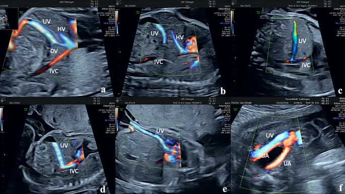
Ductus venosus flow | FMF Certification | Welcome to the Fetal Medicine  Foundation
Ductus venosus flow | FMF Certification | Welcome to the Fetal Medicine Foundation

Abnormal Waveforms In the Ductus Venosus

Ultrasound the Best #16: Ductus Venosus Waveform – WFUMB

Fetal Medicine Foundation - Ductus venosus - YouTube

Fetal ductus venosus flow assessment | Radiology Reference Article |  Radiopaedia.org
Fetal ductus venosus flow assessment | Radiology Reference Article | Radiopaedia.org

Normal ductus venosus doppler trace. | Download Scientific Diagram

Doppler Sonography of the Fetal Venous Circulation | Radiology Key

ASSESSMENT OF DUCTUS VENOSUS BY DOPPLER ULTRASOUND IN LOW RISK PREGNANCIES  OF 20-38 WEEKS GESTATION | Semantic Scholar
ASSESSMENT OF DUCTUS VENOSUS BY DOPPLER ULTRASOUND IN LOW RISK PREGNANCIES OF 20-38 WEEKS GESTATION | Semantic Scholar